Lesson 25.2: Female Reproductive System
Lesson 25.2: Female Reproductive System
Lesson Objectives
Explain how the female reproductive system develops before birth and matures during puberty.
Identify structures of the female reproductive system and their functions.
Describe how eggs are produced and how they are released from the ovaries.
Sequence the events of the menstrual cycle, and explain how hormones control the cycle.
Introduction
The female reproductive system is a collection of organs and other structures located primar- ily in the pelvic region. Most of the structures are inside the body. The female reproductive system has several functions:
producing eggs, which are female gametes
secreting female sex hormones
receiving sperm during sexual intercourse
supporting the development of a fetus
delivering a baby during birth
breastfeeding a baby after birth
During puberty, a girl develops into a sexually mature woman, capable of producing eggs and reproducing.
Sexual Development in Females
As you read in Lesson 25.1, the main differences between boys and girls at birth are their re- productive organs. Unlike males, females are not influenced by the male sex hormone testos- terone during embryonic and fetal development. This is because they lack a Y-chromosome. As a result, females do not develop male reproductive organs.
Development Before Birth
Unless an embryo is stimulated by testosterone, the reproductive organs develop into female organs, such as the ovaries and uterus. By the third month of fetal development, most of the internal female organs have formed. Immature ova, or eggs, also form in the ovary before birth. Whereas a male produces sperm throughout his lifetime (after puberty), a female produces all the eggs she will ever make before birth.
Like baby boys, baby girls are born with all their reproductive organs present but immature and unable to function. Female reproductive organs grow very little during childhood. They begin to grow rapidly and to mature during puberty.
Changes of Puberty
From Lesson 25.1, you know that puberty is the period during which humans become sexually mature. Puberty in girls differs from puberty in boys in several ways, including when it begins, how long it lasts, and the hormones involved. Girls begin puberty a year or two earlier than boys, and they complete puberty in about four years instead of six. In females, the major sex hormone is estrogen rather than testosterone.
Puberty in girls starts when the hypothalamus in the brain stimulates the pituitary gland to secrete hormones that target the ovaries. The pituitary hormones are luteinizing hormone, or LH, and follicle-stimulating hormone, or FSH. These hormones stimulate the ovary to produce estrogen.
Estrogen has many functions that you will read more about below. During puberty, estrogen promotes growth and other physical changes in females. For example, estrogen stimulates growth of the breasts and uterus. It also stimulates development of bones and contributes to the adolescent growth spurt in height. These and several other changes in females during puberty are listed in Table 25.3:
Table 25.3: Physical Changes in Females During Puberty
Changes in Reproductive Organs
Ovaries and follicles grow Uterus grows and endometrium thickens Other reproductive structures grow Menstrual cycle begins
Other Physical Changes
Breasts develop Long bones grow and mature
Pubic hair grows Underarm hair grows
Body fat increases Apocrine sweat glands develop Pelvis widens
Some of the changes involve the maturation of organs, such as ovaries, that are necessary for reproduction. Mature reproductive organs are primary sex characteristics. Other changes, such as growth of pubic hair, lead to traits that are secondary sex characteristics. One of the most significant changes in females during puberty is menarche. Menarche is the beginning of menstruation, or monthly periods, which will be discussed later.
Adolescent Growth Spurt
Females go through an adolescent growth spurt in height as boys do. However, the growth spurt in girls starts a year or two earlier and ends about three years sooner. Girls also do not grow as rapidly during their peak growth rate. Although females start the growth spurt only 2 centimeters shorter than males, on average, by the time they stop growing females are an average of 10 centimeters shorter.
Timing of Puberty
The changes of puberty usually happen in the same order for most females. The first observ- able change is typically the beginning of breast development. This happens by age 10 years in the majority of girls in the U.S. The appearance of pubic hair usually occurs next, at age
10.5 years, on average. The growth spurt in height also usually begins during the first year of puberty. During the first two years of puberty, the ovaries and uterus gradually increase in size. Menarche occurs relatively late in puberty, typically between the ages of 12 and 13 years in U.S. girls. After menarche, a female generally keeps growing for another year or two and attains her adult height by an average age of 14.5 years.
As in males, there is a wide range of ages at which particular changes of female puberty normally occur. For example, menarche may occur as early as age 8 years or as late as age
Differences in age at menarche and other changes of puberty are due to both genetic factors and environmental factors, such as diet. A female who goes through puberty earlier or later than her peers may worry that she is not developing normally. Although such variation is usually normal, she should talk with her health care provider if she has concerns.
Female Reproductive Organs
The female reproductive system is shown in Figure 25.4. Only a few of the structures are external to the body. All the main reproductive organs are internal.
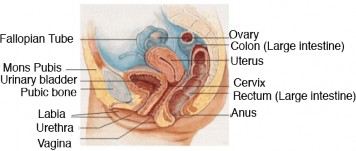
Figure 25.4: The female reproductive system. (15)
External Organs
The external female reproductive structures are referred to collectively as the vulva. They include the labia and mons pubis. The labia are the “lips” of the vulva. They protect the vagina and urethra, both of which have openings in the vulva. The mons pubis consists of fatty tissue covering the pubic bone. It protects the pubic bone and vulva from injury.
Internal Organs
The internal female reproductive organs include the vagina, uterus, fallopian tubes, and ovaries. These organs are shown from the front, without any other structures blocking them, in Figure 25.5. This makes it easier to see the shape and size of the organs and where they are located relative to one another.
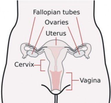
Figure 25.5: Internal female reproductive organs. (13)
The vagina is a tube-like structure about 8 to 10 centimeters long. It begins at the vulva and extends to the uterus. It has muscular walls lined with mucous membranes. The vagina has two major reproductive functions. It receives sperm during sexual intercourse, and it provides a passageway for a baby to leave the mother’s body during birth.
The uterus is a muscular organ about 7.5 centimeters long and 5 centimeters wide. It has a thick lining of tissues known as the endometrium. The lower, narrower end of the uterus is called the cervix. The uterus is where a fetus grows and develops until birth. During pregnancy, the uterus can expand dramatically to accommodate the growing baby. Muscular contractions of the uterus push the baby through the cervix during childbirth.
Extending from the upper corners of the uterus are the two Fallopian tubes. The tubes are about 7 to 14 centimeters long. Each tube reaches (but is not attached to) one of the ovaries. The ovary end of the tube has a fringelike structure (Figure 25.8) that moves with a wavelike motion.
The two ovaries are small, oval-shaped organs that lie on either side of the uterus. They are the egg-producing organs of the female reproductive system, and they contain hundreds of thousands of immature eggs. Each egg is located within a structure called a follicle. A follicle consists of the egg surrounded by special cells that protect the egg until puberty and then help the egg mature.
The Breasts
The breasts are considered secondary sex characteristics, rather than organs of reproduction. They are described here because of their role in nurturing an infant after birth. As shown in Figure 25.6, each breast contains mammary glands. The cells of mammary glands secrete milk, which drains into ducts leading to the nipple. A suckling baby squeezes the milk out of the ducts and through the nipple.
Egg Production
At birth, a female’s ovaries contain all the eggs she will ever produce. However, the eggs do not start to mature until she enters puberty. After menarche, one egg typically matures each month throughout a female’s adult years until she reaches middle adulthood.
Oogenesis
Oogenesis is the process of producing eggs in the ovary. Eggs are haploid cells, having half the number of chromosomes of other cells in the body, which are diploid cells. Like sperm, eggs must be haploid in order for sexual reproduction to result in diploid offspring. Like spermatogenesis, oogenesis occurs in several steps that involve different types of cells. The steps of oogenesis are listed in Table 25.4.
Oogenesis begins when an oogonium with the diploid number of chromosomes undergoes mitosis to form primary oocytes, also with the diploid number. It proceeds as a primary oocyte undergoes the first cell division of meiosis to form secondary oocytes with the haploid number of chromosomes. A secondary oocyte undergoes the second meiotic cell division to form a haploid ovum if it is fertilized by a sperm.
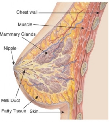
Figure 25.6: Cross-section of a human breast. (14)
Table 25.4: Oogenesis and Cell Division
|
Type of Cell |
Number of Chromosomes |
Process |
|
Oogonium |
Diploid |
Mitosis |
|
Primary oocyte |
Diploid |
Meiosis 1 |
|
Secondary oocyte |
Haploid |
Meiosis 2 |
|
Ovum (mature egg) |
Haploid |
Fertilization |
Oogenesis begins with oogonia (singular, oogonium), which are the immature eggs that form in the ovaries before birth. Oogonia are diploid cells and equivalent to spermatogonia in males. By about the fifth month of fetal development, the ovaries contain about seven million oogonia.
Over the next few months, oogonia undergo mitosis, forming cells called primary oocytes. Primary oocytes are also diploid cells. Before birth, primary oocytes begin the first division of meiosis, but they do not complete it until long after birth. At birth, the average female has about two million primary oocytes in her ovaries. Throughout childhood, the number of oocytes falls as they deteriorate and disappear. By puberty, there are only about 300,000 to 400,000 primary oocytes left in the average girl’s ovaries.
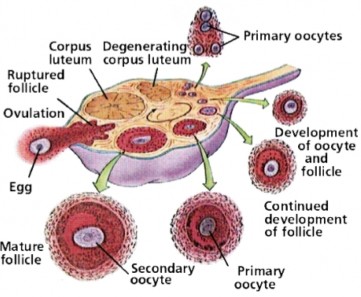
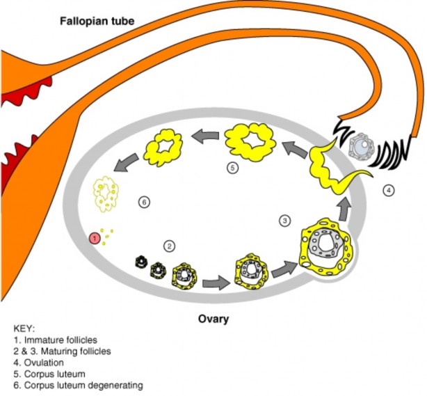
Maturation of a Follicle
Beginning in puberty, each month one of the follicles starts to mature (Figure 25.7). The primary oocyte in the follicle resumes meiosis and divides to form a secondary oocyte and a smaller cell, called a polar body. Both the secondary oocyte and polar body are haploid cells. The secondary oocyte has most of the cytoplasm from the original cell and is much larger than the polar body. The polar body disintegrates and disappears from the ovary.
Ovulation
Ovulation is the release of a secondary oocyte by the ovary. Ovulation occurs every 28 days, on average, in a sexually mature female, but may range normally from 24 to 36 days. As shown in Figure 25.7, during ovulation a secondary oocyte bursts out of its follicle and through the ovary wall to enter the abdominal cavity.
Each month only one of the ovaries matures a follicle and releases an egg. Which ovary matures a follicle in a given month? Scientists say that it appears to be random.
After the secondary oocyte leaves the ovary, it is swept into the Fallopian tube by the waving, fringelike end. This is illustrated in Figure 25.8. Tiny hairlike projections, called cilia, line the tube and help move the oocyte through to the uterus. If the secondary oocyte is fertilized by a sperm as it is passing through the Fallopian tube, it divides to form a mature egg and
a polar body, finishing meiosis. (As before, the polar body contains very little cytoplasm and disintegrates.) If the secondary oocyte is not fertilized, it passes into the uterus as an immature egg.
Menstrual Cycle and Menstruation
Ovulation is part of the menstrual cycle, which occurs each month in a sexually mature female. Another part of the cycle is menstruation. Menstruation is the process in which blood and other tissues are shed from the uterus and leave the body through the vagina. It is also called a menstrual period, or menses. The menstrual cycle is sometimes divided into two cycles, called the ovarian cycle and the uterine cycle. The ovarian cycle includes the events that occur in the ovary. The uterine cycle refer to the events that occur in the uterus. The two cycles are closely related, so here they are described together and referred to jointly as the menstrual cycle.
Phases of the Menstrual Cycle
The phases of the menstrual cycle are summarized in Table 25.5. The cycle begins with the menstrual phase, which typically lasts from one to four days. This is when menstruation occurs. During the menstrual phase, arteries that supply the endometrium of the uterus constrict and break. Gradually, blood and endometrial tissues detach from the inside of the uterus and pass from the uterus to the vagina and then out of the body. If there is an immature egg in the uterus, it passes out of the body with the menstrual flow.
The menstrual cycle (as shown in Table 25.5) includes an ovarian and a uterine cycle. Events in the ovarian cycle include maturation of a follicle, release of an egg, and formation of the corpus luteum. Events in the uterine cycle include menstruation, development of the endometrium, and thickening of the endometrium in preparation for an egg.
Table 25.5: The Phases of the Menstrual Cycle
Name of Phase Days Events
Menstrual Phase 1–4 Menstruation occurs
Follicular Phase 5–13 Follicle matures En- dometrium develops
Ovulation 14 Ovary releases egg
Luteal Phase 15–28 Follicle becomes corpus luteum Endometrium prepares for egg
The next phase of the cycle is called the follicular phase. After menstruation, the en- dometrium in the uterus begins to build up again. At the same time, several follicles start maturing in the ovary. Only one of these maturing follicles will complete maturation. The rest will eventually deteriorate and disappear. By the middle of the menstrual cycle, around day 14, the remaining mature follicle releases its oocyte from the ovary in the process of ovulation.
Following ovulation, the luteal phase begins. During the luteal phase, the endometrium of the uterus continues to prepare for a fertilized egg. For example, it becomes thicker and develops more blood vessels. At the same time, the mature follicle that just released its egg develops into a structure called a corpus luteum (Figure 25.8).
If the egg is fertilized and implants, or embeds itself, in the endometrium of the uterus, the endometrium will be maintained and help nourish it. If the egg is not fertilized, the endometrium will break down, leading to menstruation. This begins a new cycle.
The events of the menstrual cycle always occur in the same sequence, but their timing may vary considerably. There is a great deal of normal variation in the length of the overall cycle and of the individual phases. Variation may occur from one female to another and also from one cycle to the next for a given female.
Some females have symptoms—such as bloating, abdominal cramps, and mood swings—for several days before or during menstruation each month. If the symptoms are severe enough to interfere with daily life, the condition is called premenstrual syndrome, or PMS. Symptoms of PMS often can be helped with medications or lifestyle changes.
Role of Hormones
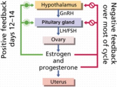
The same hormones that control female puberty and oogenesis also control the menstrual cycle: estrogen, LH, and FSH. Estrogen controls the secretion of the two pituitary hormones by acting on the hypothalamus, which controls the pituitary gland. This is shown in Fig- ure 25.9. When the estrogen level rises in the blood, it stimulates the pituitary (via the hypothalamus) to secrete more or less LH and FSH.
In negative feedback, rising levels of hormones feedback to the hypothalamus and pituitary gland to decrease production of the hormones. In positive feedback, rising levels of hormones feedback to increase hormone production. During most of the menstrual cycle, estrogen and progesterone provide negative feedback to the hypothalamus and pituitary gland. This keeps their levels more or less constant. During days 12–14, however, estrogen provides positive feedback to the hypothalamus and pituitary gland. This causes a rapid rise in the production of estrogen by the ovary and leads to ovulation.
Another hormone involved in the menstrual cycle is progesterone. The word ”progesterone” literally means “pro-gestational hormone.” Progesterone is a hormone that promotes ges- tation, or the carrying of a fetus. The function of progesterone in the menstrual cycle is to maintain the endometrium of the uterus.
Change in the levels of these four hormones (estrogen, LH, FSH, and progesterone) occur during the menstrual cycle (Figure 25.10). After menstruation occurs, estrogen secreted by the ovaries increases. This causes the endometrium of the uterus to thicken. FSH from the pituitary stimulates follicles in the ovary to mature. The maturing follicles produce estrogen, and the level of estrogen in the blood rises. When estrogen reaches a high level in the blood,
it stimulates the pituitary gland to release a surge of LH. The spike in LH stimulates the one remaining mature follicle to burst open and release its oocyte.
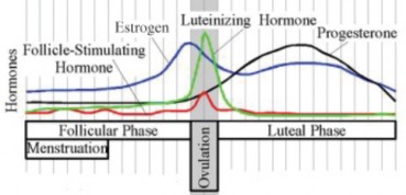
Figure 25.10: This graph shows how hormone levels change during the menstrual cycle. (5)
During the first half of the cycle, negative feedback keeps levels of FSH, LH, estrogen, and progesterone relatively stable. During ovulation, positive feedback causes a burst of FSH, LH, and estrogen. During the second half of the cycle, progesterone rises as the corpus luteum in the ovary matures and produces this hormone. Negative feedback helps keep levels of the other three hormones fairly constant
After the oocyte is released, LH stimulates the mature follicle to develop into a corpus luteum. The corpus luteum then starts secreting progesterone, which maintains the endometrium of the uterus. What happens next depends on whether the egg has been fertilized.
f the egg has been fertilized, it will soon start producing a hormone that helps maintain the corpus luteum. As a result, the corpus luteum will continue producing progesterone and maintain the endometrium.
If the egg has not been fertilized, the corpus luteum will disintegrate and stop producing progesterone. Without progesterone, the endometrium will break down, detach from the uterus, and pass out of the body during menstruation.
Menopause
For most women in the U.S., the menstrual cycle continues into their forties. Then it gradually becomes more and more irregular until it finally stops altogether, generally by their early fifties. Menopause occurs when a woman has gone through 12 consecutive months without a menstrual period. She can no longer reproduce because her ovaries no longer produce eggs.
The cause of menopause is a natural decline in estrogen secretion by the ovaries as a woman ages. It may take from several months to a few years for her body to adjust to the drop in estrogen. During this time, she may experience hot flashes, mood swings, and other symptoms.
Lesson Summary
The female reproductive system forms before birth but does not become capable of reproduction until it matures during puberty.
The female reproductive system includes organs and other structures that produce and release eggs, secrete female sex hormones, and enable the development and birth of a fetus.
Immature eggs form in the ovaries before birth. Each month, starting in puberty, one egg matures and is released from the ovary.
The menstrual cycle includes events that take place in the ovary, such as ovulation, and changes in the uterus, including menstruation. The menstrual cycle controlled by the hormones estrogen, progesterone, LH, and FSH.
Review Questions
Further Reading / Supplemental Links
List three functions of the female reproductive system.
State two ways that puberty differs in girls and boys.
Describe the uterus and its functions in reproduction.
What is ovulation and when does it occur?
Tara is 13 and worried that she may not be developing normally. She began developing breasts about six months ago but still has not had her first menstrual period. Should
she be concerned? Explain your answer.
Explain how blockage of both Fallopian tubes would affect a woman’s ability to repro- duce naturally.
Create a timeline showing the steps in which an oogonium develops into a mature egg.
Explain the roles of estrogen, LH, and FSH in the menstrual cycle.
Stanley, Deborah, Sexual Health Information for Teens. Omnigraphics, 2003.
Walker, Pam and Wood, Elaine, Understanding the Human Body: The Reproductive System. Lucent Books, 2002.
http://en.wikibooks.org/wiki/Human_Physiology/The_female_reproductive_system
http://www.kidshealth.org/parent/general/body_basics/female_reproductive_ system.html
http://www.kidshealth.org/teen/sexual_health/changing_body/female_repro.
http://www.medicalook.com/human_anatomy/systems/Female_reproductive_system. html
http://www.merck.com/mmhe/sec22/ch241/ch241a.html
Vocabulary
adolescent growth spurt Rapid growth in height seen during puberty.
corpus luteum Formed in the ovary from the ruptured follicle after ovulation; if the egg is not fertilized by a sperm, the corpus luteum degenerates and virtually disappears from the ovary; produces progesterone.
egg (ova) Female gamete, or sex cell, which is necessary for reproduction; haploid.
estrogen Major female sex hormone.
Fallopian tube Tube which accepts oocyte after ovulation; site of fertilization; attached to uterus.
female reproductive system System with several major functions: producing eggs, se- creting female sex hormones, receiving sperm during sexual intercourse, supporting the development of a fetus, delivering a baby during birth, and breastfeeding a baby after birth.
follicle Structure in which each egg is located; consists of the egg surrounded by special cells that protect the egg until puberty and then help the egg mature.
follicle-stimulating hormone (FSH) Hormone that stimulates the ovary to produce es- trogen.
luteinizing hormone (LH) The main pituitary hormone responsible for puberty in fe- males; stimulates the ovary to produce estrogen.
menarche The beginning of menstruation, or monthly periods.
menopause When a woman has gone through 12 consecutive months without a menstrual period; she can no longer reproduce because her ovaries no longer produce eggs.
menstruation The process in which blood and other tissues are shed from the uterus and leave the body through the vagina; also called a menstrual period, or menses.
oogenesis The process of producing eggs in the ovary.
ovary Small, oval-shaped organs that lie on either side of the uterus; the egg-producing organs of the female reproductive system; contain hundreds of thousands of immature eggs.
ovulation The release of a secondary oocyte by the ovary; occurs every 28 days, on average.
progesterone A hormone that promotes gestation, or the carrying of a fetus; also main- tains the endometrium of the uterus.
uterus A muscular organ where a fetus grows and develops until birth; has a thick lining of tissues known as the endometrium; the lower, narrower end of the uterus is called the cervix.
vulva The external female reproductive structures; includes the labia and mons pubis.
Points to Consider
If an egg is fertilized by a sperm and implants in the uterus, the endometrium helps support and nourish it. However, the new organism soon needs more nutrients than the endometrium can provide. It needs to obtain nutrients from the mother’s blood. How does this happen?
What structures are involved with pregnancy? When do they develop?
- Log in or register to post comments
- Email this page