Lesson 25.3: Fertilization, Gestation, and Devel- opment
Lesson 25.3: Fertilization, Gestation, and Devel- opment
Lesson Objectives
Explain how fertilization, cleavage, and implantation lead to the formation of an em- bryo.
Describe how the embryo forms specialized cells and organs through the processes of gastrulation, differentiation, and organogenesis.
Identify major events in the growth and development of the fetus.
Explain how the placenta provides the fetus with oxygen and nutrients and eliminates fetal wastes.
Describe how an expectant mother can help her fetus grow and develop normally, and summarize the events of childbirth.
Sequence milestones in growth and development from infancy through adolescence.
Describe the life stages of early and middle adulthood and old age, and explain why aging occurs.
Introduction
Sexual reproduction begins when an egg is fertilized by a sperm and implants in the uterus. Following these events, the remainder of growth and development before birth is divided into two main stages. The first stage is the embryonic stage, which lasts about two months. This is followed by the fetal stage, which lasts for another seven months until birth.
Fertilization, Cleavage, and Implantation
A day or two after an ovary releases an egg, the egg may unite with a sperm. However, before it becomes an embryo, it must go through other processes. These processes include cleavage and implantation.
Fertilization
Fertilization is the union of a sperm and an egg. Recall that a sperm is a male gamete and an egg is a female gamete. Each gamete is a haploid cell. When the two cells unite during fertilization, they form a diploid cell, called a zygote.
Fertilization generally occurs in a Fallopian tube. After sperm are deposited in the vagina during sexual intercourse, they “swim” through the cervix and uterus and into a Fallopian tube. Although millions of sperm are deposited, only a few hundred are likely to reach the egg. A sperm about to penetrate an egg is shown in Figure 25.11. When a sperm finally breaks through the egg’s cell membrane, it sets off a reaction that prevents other sperm from entering. The entry of the sperm also triggers the egg to complete the second meiotic division that began before ovulation.
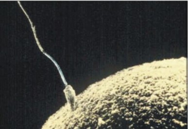
Figure 25.11: Human sperm and egg. (19)
After the sperm penetrates the egg, its tail falls off, and its nucleus fuses with the nucleus of the egg. The resulting zygote contains all the chromosomes needed for a new individual. Half the chromosomes are from the egg, and half are from the sperm.
Cleavage
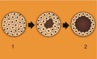
The zygote spends the next few days traveling down the Fallopian tube. As it travels, it divides by mitosis several times to form a ball of cells called a morula. The cell divisions, which are called cleavage, increase the number of cells but not their overall size. More cell divisions occur, and soon a fluid-filled cavity forms inside the ball of cells. At this stage, the ball of cells is called a blastocyst. The process of blastocyst formation is shown in Figure 25.12.
The cells of the blastocyst form an inner and an outer cell layer. This is apparent in Figure
The inner layer of cells is called the embryoblast. This layer of cells will soon develop into an embryo. The outer layer of cells is called the trophoblast. This layer will develop into other structures, including the placenta, which you will read more about below.

Implantation
The blastocyst continues the trip down the Fallopian tube and reaches the uterus about four or five days after fertilization. When the outer cells of the blastocyst contact cells lining the uterus, the blastocyst embeds in the lining. The process of embedding is called implantation. It generally occurs about a week after fertilization. Once implantation occurs, the blastocyst is called an embryo.
Growth and Development of the Embryo
An embryo is a developing human being from the time of implantation through the first eight weeks after fertilization. During this time, the embryo grows in size and undergoes three processes: gastrulation, differentiation, and organogenesis.
Gastrulation
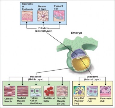
Gastrulation is the development of different layers of cells in the embryo. It generally occurs during the second week after fertilization. During gastrulation, cells of the embryo migrate to form three distinct cell layers: the ectoderm, mesoderm, and endoderm. These layers are shown in Figure 25.14. Each layer will eventually develop into certain types of tissues and cells in the body.
Ectoderm—Forms tissues that cover the outer body; develops into cells such as nerves, skin, hair, and nails.
Mesoderm—Forms tissues that provide movement and support; develops into cells such as muscles, bones, teeth, and blood.
Endoderm—Forms tissues involved in digestion and breathing; develops into cells such as lungs, liver, pancreas, and gall bladder.
Differentiation and Organogenesis
During the third week after fertilization, the embryo begins to undergo cellular differenti- ation. Differentiation is the process by which unspecialized cells become specialized into one of the many different types of cells that make up the body. During differentiation, certain genes are turned on, or activated, while other genes are switched off, or inactivated. As a result of this process, cells develop specific structures and abilities that suit them for their specialized roles in the body. Several examples of specialized cells are shown in Figure 25.14, along with the cell layers from which they develop.
Differentiation of cells leads to the development of specific organs within the three cell layers. This is called organogenesis. All the major organs begin to form during the remaining
weeks of embryonic development. A few of the developments that occur in weeks 4 through 8 are listed below.
Embryonic Development During Weeks 4-8
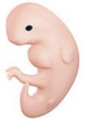
Pictured above is a 4-week-old embryo.
At Week 4
Heart begins to beat.
Arm buds appear.
Liver, pancreas, and gall bladder start to form.
Spleen appears.
At Week 5
Eyes start to form.
Leg buds appear.
Hands appear as paddles.
Blood begins to circulate.
Facial features start to develop.
At Week 6
Lungs start to form.
Fingers and toes form.
At Week 7
Hair follicles start to form.
Elbows and toes are visible.
At Week 8
Facial features look more human.
External ear begins to take shape.
As the embryo develops, it also grows in size. By the eighth week of development, the embryo is about 30 millimeters long. It may also have begun to move.
Growth and Development of the Fetus
From week 8 until birth, the developing individual is referred to as a fetus. In humans, birth typically occurs 38 weeks after fertilization, so the fetal period lasts about 30 weeks. During this time, the organs that formed during the embryonic period go through further develop- ment. The fetus also grows in overall body size. For a detailed animation of the growth and development of the fetus see http://www.youtube.com/watch?v=aR-Qa_LD2m4& feature=related.
Weeks 8 to 15
During the fetus’s early weeks, reproductive organs develop along either male or female lines. The liver starts producing red blood cells, and tooth buds appear. The fetus becomes more human in appearance, with well-formed facial features. The eyelids form but remain closed until later in fetal development. The muscles and bones develop, and the fetus is very active. It can make a fist and move its arms and legs. It also hiccups, stretches, and yawns. The first measurable brain activity occurs around the 12th week. By the end of the 15th week, the fetus is about 15 centimeters long.
Weeks 16 to 26
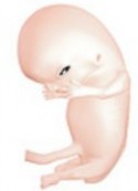
A fetus at 18-weeks after fertilization is shown in Figure ??. At this stage, the brain is developing rapidly, and it starts to take control of some body functions. The alveoli (air sacs) in the lungs also develop, making gas exchange possible, although the lungs are still immature. Most of the internal components of the eyes and ears form and develop at this time. There is more muscle development, as well, and the fetus is more active than ever. The mother usually starts to feel fetal movement during this stage.
Fine hair called lanugo grows and covers the fetus’s body by the end of this stage. Eyebrows, eyelashes, and nails also appear, and the eyelids begin to open and close. By the end of week 26, the fetus is about 38 centimeters long and weighs about 1.2 kilograms.
Weeks 27 to 38
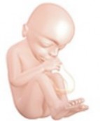
During the final weeks of growth and development, the amount of body fat rapidly increases. Bones develop fully, although they are still soft and pliable. Most of the lanugo disappears, and head hair becomes coarser and thicker. Fingernails grow beyond the end of the fingertips. In the brain, connections form that allow the input of sensations. Starting around week 30, the brain is continuously active. By the 38th week, the fetus is fully developed and ready to be born. A 38-week fetus normally ranges from 36 to 51 centimeters in length and weighs between 2.7 and 4.6 kilograms. A 38-week-old fetus is shown in Figure ??.
Sometimes fetuses are born earlier than 38 weeks. After 35 weeks, the fetus is considered “full-term,” which means that it is developed enough for life outside the mother. Fetuses born before 35 weeks are likely to have health problems due to their immaturity, although many are able to survive with medical help. The less time a fetus spends developing in the uterus before it is born, the less likely it is to survive after birth. Fetuses born before 25 weeks rarely survive.
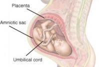
Placenta and Related Structures
The placenta is a temporary organ in which nutrients and wastes are exchanged between the mother and the embryo or fetus. The placenta begins to form in the second week after fertilization. It continues to develop and grow to meet the needs of the growing fetus. A fully developed placenta, like the one in Figure ??, is made up of a large mass of blood vessels from both the mother and fetus. The maternal and fetal vessels are close together but separated by empty space. This allows the mother’s and fetus’s blood to exchange substances without actually mixing.
How the Placenta Works
Blood from the mother enters the maternal blood vessels of the placenta under pressure, forcing the blood into the empty spaces. When the mother’s blood contacts the fetal blood vessels, gases are exchanged. Oxygen from the mother’s blood is exchanged with carbon dioxide from the fetus’s blood. A release of pressure brings the mother’s blood back from the placenta and into her veins.
The fetus is connected to the placenta through the umbilical cord, a tube that contains two arteries and a vein. Blood from the fetus enters the placenta through the umbilical arteries, exchanges gases with the mother’s blood, and travels back to the fetus through the umbilical vein.
In addition to gas exchange, the placenta transfers nutrients, hormones, and other needed substances from the mother’s blood to the fetus’s blood. The placenta also filters many harmful substances out of the mother’s blood so they are not transferred to the fetus. In addition, the placenta secretes hormones that maintain the corpus luteum in the mother’s ovary. Recall that the corpus luteum secretes progesterone, which is needed to keep the endometrium of the uterus from breaking down.
Amniotic Sac and Fluid
Attached to the placenta is the amniotic sac, which surrounds and protects the embryo or fetus. It begins to form in the second week after fertilization. It soon fills with water and dissolved substances to form amniotic fluid. The fluid allows the fetus to move freely until the fetus grows to fill most of the available space. The fluid also cushions the fetus and helps protect it from injury.
Pregnancy and Childbirth
Pregnancy is the carrying of one or more offspring from fertilization until birth. It is the development of a fetus from the expectant mother’s point of view. A woman is likely to first suspect she is pregnant when she misses a menstrual period. As you just read, hormones secreted by the placenta maintain the endometrium of the uterus. This prevents menstruation from occurring once pregnancy begins.
The pregnant mother plays a critical role throughout the embryonic and fetal periods. She must provide all the nutrients and other substances needed for normal growth and develop- ment. Therefore, it is important for the expectant mother to take good care of her health during pregnancy for the sake of her baby as well as herself. Most importantly, the mother needs to avoid toxic substances and take in adequate nutrients.
Avoiding Toxins
Unfortunately the placenta cannot protect the developing embryo or fetus from all harmful substances in the mother’s blood. Some harmful substances can cross the placenta from the mother’s blood and damage the embryo or fetus, including:
Alcohol
Chemicals in tobacco smoke
Aspirin
Thalidomide (a prescription drug)
Heroin
Cocaine
These and other substances can cause birth defects. For example, if a pregnant woman drinks alcohol, it can cause variety of birth defects that are collectively called fetal alcohol syndrome. A baby with fetal alcohol syndrome is shown in Figure 25.15. The defects include facial abnormalities, stunted growth, and mental retardation.
Alcohol and some other toxins can damage the developing brain at any time before birth because the brain continues to develop and grow rapidly throughout pregnancy. However, in general, birth defects are likely to be more severe when exposure to toxins occurs during the embryonic period. This is because the embryo is undergoing organogenesis. Any disruption of normal development during this early period is likely to have a greater impact on the organism than later in pregnancy, when the organs are already formed. Although exposure to toxins at later stages of development may do less damage, an expectant mother should try to avoid toxins throughout her pregnancy.
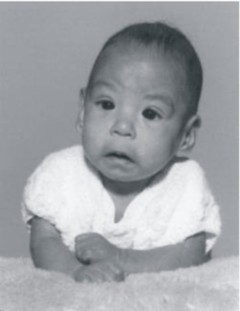
Figure 25.15: Baby with fetal alcohol syndrome. (11)
Taking in Nutrients
The fetus depends completely on the mother for its nutrient needs. As a result, most nutrients are needed in greater amounts by a pregnant woman than a woman who is not pregnant. Some nutrients are especially important for embryonic or fetal development.
Folic acid (vitamin B9) is needed for normal development of the spinal cord. Inadequate folic acid intake can lead to spina bifida, a serious birth defect.
Calcium is needed for normal development of bones and teeth.
Iron is needed for the proper formation of red blood cells.
Omega-3 fatty acids are important for normal development of nerve cells.
If an expectant mother eats a balance of foods from the different food groups, this diet will help ensure adequate nutrients for the fetus. Because needs for some nutrients are so high, nutrient supplements are usually recommended during pregnancy. Supplements formulated for pregnant women help supply adequate amounts of folic acid and other nutrients needed for normal growth and development of the fetus.
Childbirth
Near the time of birth, the amniotic sac breaks in a gush of fluid. Within 24 hours of the amniotic sac breaking, labor usually begins. Labor involves contractions of the muscular walls of the uterus. The contractions are stimulated by the release of the pituitary hormone oxytocin. The contractions cause the cervix to widen and the passage through the cervix to dilate, or open. The contractions become closer and stronger, and the cervix gradually becomes more dilated. This may take hours or even days. When the cervix is dilated to about 10 centimeters, the baby begins to move through cervix and into the vagina.
At this point, the mother begins pushing to aid in the birth of the baby. This part of labor is generally shorter. The fetus usually emerges head first. Within seconds of birth, the umbilical cord is cut. Without this connection to the placenta, the baby cannot exchange carbon dioxide, which quickly builds up in the baby’s blood. This stimulates the brain to trigger breathing and the newborn takes its first breath. Generally within half an hour or less of the birth of the baby, contractions of the uterus force the placenta and any remaining amniotic tissues from the mother’s body.
By birth, a fetus has a large head relative to its body size, because the brain is more developed than any other organ. Some areas of the skull have not yet been converted to hard bone, allowing the fetus’s head to change shape somewhat to fit through the cervix during birth. The head returns to its normal shape shortly after birth.
Infancy, Childhood, and Adolescence
For the first year after birth, a baby is called an infant. Early childhood begins at age two, when a child may be referred to as a toddler. Childhood continues until adolescence, which generally coincides with the teen years. Adolescence is the period of transition into adulthood.
Infancy
Infancy is defined as the first year of life after birth. For the first month after birth, an infant is called a newborn. A newborn has a distinctive appearance. The head is very large, and the arms and legs are relatively short. The shoulders and hips are narrow, and the abdomen protrudes slightly. Many newborns still have lanugo on some areas of their body, but this usually disappears within a few weeks after birth. Head hair can vary from almost no hair to a full head of hair. The stub of the umbilical cord remains for a few weeks, until it dries up and falls off, forming the navel.
Infants are born with certain abilities already developed. For example, they have a well developed sense of smell. They can also communicate their needs by crying when they are hungry, uncomfortable, bored, or lonely. During their first year, they develop many other abilities:
By 6 weeks after birth, babies typically start smiling (Figure 25.16) and making vocal sounds.
By 6 months, they spend a lot of time babbling. They have also learned to sit and are starting to crawl.
By 12 months, they are saying their first words. They can stand with help and may have started to walk.
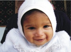
Figure 25.16: Six-week-old baby’s first smile. (6)
Infancy is the period of most rapid growth after birth. Growth during infancy is even faster than growth during puberty. By the end of the first year, the average baby is twice the length it was at birth and three times its birth weight. Infancy is also the period when most of the deciduous, or “baby,” teeth erupt. The front teeth erupt first, usually starting around six months after birth. There are 20 deciduous teeth altogether, and they continue to erupt until about three years of age.
Newborns need about 18 hours of sleep each day. They usually sleep in long naps throughout the day and night. As infants get older, they need less sleep. They also start to sleep through the night and just take short naps during the day. When newborns aren’t sleeping, they are usually feeding. Breastfeeding is the recommended method of feeding infants. Breast milk is generally supplemented by other foods by the end of the first year.
Childhood
A toddler is a young child who is learning to walk, or “toddle.” This is the second stage of development after infancy. It generally refers to children between the ages of 1 and 3 years. During this stage, children not only learn to walk steadily but also develop other motor skills. By the end of the third year, most children can run, walk up steps, and climb onto chairs. They can feed and dress themselves with help. They can also manipulate small objects and hold a crayon and scribble with it. They have learned dozens of words and are speaking in simple sentences. Most children are also toilet trained by the end of the third year.
Growth is still relatively rapid during the toddler years but slowing down. By the time children are five years old, their height is increasing by only about 5 percent per year, compared with 100 percent per year in the first year of life. By age five, children are able to carry on conversations, recognize letters and words, and tie their shoe laces. Five-year olds can use a pencil to trace letters and other shapes (Figure 25.17). They also may be learning to ride a bicycle, swim, swing a bat, or kick a ball.
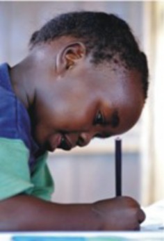
Figure 25.17: A five-year old using a pencil to trace shapes. (9)
By age six, most children begin losing their deciduous teeth, and their permanent teeth erupt to replace them. This continues until about age 12. Other important changes of older childhood include the transition from home to school. At school, children not only acquire academic skills such as reading, but also interact more with their peers. They form friendships and are likely to have “best” friends. Older children continue to grow slowly until they start the adolescent growth spurt during puberty.
Adolescence
Adolescence is the period of life between the beginning of puberty and adulthood. You learned about the physical changes of puberty earlier in this chapter. Adolescence is also a time of significant mental, emotional, and social changes. For example, during adolescence, teens develop more advanced mental abilities, including the ability to think abstractly. They also try to establish an identity, or sense of self. In the process, they may try to become more independent from their parents. They may also challenge authority and push limits.
Emotionally, adolescence may be a time of upheaval. Shifting hormone levels may cause mood swings at a time when many adolescents are still learning how to manage their emo- tions. One of the most important social changes of adolescence is the increased importance of peers. Teens spend much more time with their friends and other peers than younger children do, and they are generally greatly influenced by them. Young people may also start to develop intimate relationships during adolescence.
Adulthood and Old Age
The development of intimacy is considered to be a major goal of the stage of life referred to as young adulthood. Other stages of adulthood include middle adulthood and old age. Each stage is associated with particular goals and health concerns.
When Does Adulthood Start?
The age at which adulthood starts may vary from about age 17 to 21 years, depending on how adulthood is defined. A person may be physically mature by age 17 but not considered legally mature until an older age. For example, in the U.S., individuals cannot assume adult responsibilities, such as voting and joining the armed forces, until they are 18 years old. They cannot exercise certain adult rights, such as buying and using alcohol, until they are 21.
Early and Middle Adulthood
Early adulthood may be defined as the stage of life from the start of adulthood through age 34 years. During early adulthood, people generally learn how to form intimate relationships, both in friendship and love. Many people become engaged or marry during this time. Young adults may also be involved in completing their education and becoming established in a career or the workforce. Health problems in most young adults are minor. The most common causes of death are due to violence: homicides, car crashes, and suicides.
Middle adulthood may be defined as the stage of life from age 35 through 64 years. During this stage, most people raise a family (if they are going to) and strive to attain career goals. They are more likely to become involved in their community.
During middle adulthood, people start showing physical signs of aging, such as wrinkled skin and gray hair. Vision, strength, reaction time, and overall fitness also typically decline during middle adulthood. At the same time, health problems tend to increase. Diseases such as type 2 diabetes, cardiovascular disease, and many types of cancer are often diagnosed during this stage of life, especially in people who are overweight or obese. The risk of being diagnosed with diseases such as these increases throughout middle adulthood. These diseases are also the chief causes of death of middle adults.
Old Age
Old age may be defined as the stage from age 65 until death. During this stage, most people retire from work and no longer have the major responsibility of caring for others. Physically, older adults tend to have a decline in stamina, strength, reflex time, and the senses.
Other physical changes that occur in old age include a decrease in:
heart output
kidney function
lung capacity
number of brain cells
Because the immune system also becomes less efficient with age, older adults are increas- ingly susceptible to serious illnesses such as cancer, cardiovascular disease, and pneumonia. Osteoporosis, or loss of bone density, is also common in older adults, particularly in females. Mental deterioration may occur, as well, especially in people with Alzheimer’s disease and certain other diseases. Otherwise, intelligence tends to remain stable throughout adulthood and into old age.
Why does aging occur? Why does the body decline in function as people grow old? There are at least two reasons. One reason is that cells are programmed to divide a set number of times. After that, they can no longer divide, so they die out. Another reason is that DNA becomes increasingly damaged through time due to mutagens in the environment. Eventually, the damage accumulates to a point where cells can no longer divide. Most physical changes associated with aging may be due to a combination of both processes.
Lesson Summary
Fertilization is the union of a sperm cell and an egg cell that forms a zygote. The zygote undergoes many cell divisions before it implants in the lining of the uterus.
The embryonic stage begins with implantation. An embryo forms three distinct cell layers, and each layer develops into different types of cells and organs.
The fetal stage begins about two months after fertilization and continues until birth. During this stage, the organs grow and develop and the fetus grows in size.
The placenta allows nutrients and wastes to be exchanged between the mother and fetus. The fetus is connected to the placenta through the umbilical cord.
A pregnant woman should avoid toxins and take in adequate nutrients for normal fetal growth and development. During childbirth, the fetus is pushed through the cervix and out of the body through the vagina.
Growth and development are most rapid during infancy and slower throughout the rest of childhood until adolescence. Adolescence involves mental, emotional, and social changes in addition to the physical changes of puberty.
During early adulthood, people form intimate relationships and start careers. Serious health problems start showing up in middle adulthood and old age. Aging occurs as cells lose their ability to divide.
Review Questions
Further Reading / Supplemental Links
Describe what happens during fertilization.
How does gastrulation change an embryo?
Identify three events that occur as a fetus grows and develops.
Explain the role of the placenta in fetal development.
Why is an embryo generally more susceptible than a fetus to damage by toxins in the mother’s blood?
Why is the umbilical cord cut before a newborn has started to breathe on its own?
Create a timeline of growth and development from infancy through adolescence.
Explain why aging occurs.
Brynie, Faith Hickman, 101 Questions About Reproduction. 21st Century, 2004.
Stanley, Deborah, Sexual Health Information for Teens. Omnigraphics, 2003.
http://estrellamountain.edu/faculty/farabee/biobk/BioBookREPROD.html
http://www.cdc.gov/nchs/fastats/deaths.htm
http://www.keepkidshealthy.com/growthcharts/
http://en.wikibooks.org/wiki/Human_Physiology/Development:_birth_through_ death
http://www.merck.com/mmhe/sec22/ch257/ch257a.html
http://www.merck.com/mmhe/sec22/ch260/ch260a.html
http://www.visembryo.com/baby/index.html
Vocabulary
adolescence The period of life between the beginning of puberty and adulthood.
amniotic fluid Fluid that allows the fetus to move freely within the amniotic sac; also cushions the fetus and helps protect it from injury.
blastocyst The ball of cells that contains a fluid filled cavity and distinct layers; forms from the morula.
cleavage The initial cell divisions which increase the number of cells but not their overall size.
differentiation The process by which unspecialized cells become specialized into one of the many different types of cells that make up the body.
ectoderm Cell layer of the embryo that forms tissues that cover the outer body; develops into cells such as nerves, skin, hair, and nails.
embryo A developing human being from the time of implantation through the first eight weeks after fertilization.
embryoblast Inner layer of cells of the blastocyst; develops into an embryo.
endoderm Cell layer of the embryo that forms tissues involved in digestion and breathing; develops into cells such as lungs, liver, pancreas, and gall bladder.
fertilization The union of a sperm and an egg. When the two cells unite during fertiliza- tion, they form a diploid cell, called a zygote.
fetus The developing individual from week 8 until birth.
gastrulation The development of different layers of cells in the embryo; generally occurs during the second week after fertilization.
implantation The embedding of the blastocyst in the lining of the uterus; occurs about a week after fertilization. Once implantation occurs, the blastocyst is called an embryo.
infancy The first year of life after birth.
mesoderm Cell layer of the embryo that forms tissues that provide movement and support; develops into cells such as muscles, bones, teeth, and blood.
morula Initial ball of cells formed the first few days after fertilization; formed within a fallopian tube.
organogenesis The development of specific organs within the three cell layers.
placenta A temporary organ in which nutrients and wastes are exchanged between the mother and the embryo or fetus.
pregnancy The carrying of one or more offspring from fertilization until birth.
trophoblast Outer layer of cells within the blastocyst; will develop into structures which includes the placenta.
Points to Consider
Many diseases become more common as people age, but some diseases are more com- mon in adolescents and young adults, including sexually transmitted diseases (STDs). What are examples of STDs?
How common are STDs in teens and young adults?
Why are STDs more common during these two stages of life?
- Log in or register to post comments
- Email this page