Lesson 1.3: Tools and Techniques
Lesson 1.3: Tools and Techniques
Lesson Objectives
Identify the units of measurement that scientists use.
Contrast light microscopes and electron microscopes.
Identify three items that are common to science labs.
Outline the importance of mathematics to scientific research.
Outline what students and researchers can do to stay safe while working in the lab.
Introduction
Scientists need to know they are talking the same language when it comes to measurements and analysis of data. Therefore a “standard language of measurement” called the SI sys- tem is used in scientific research. Other standard procedures and techniques are carried out so that scientists from around the world can understand what was done to get to a particular conclusion. These involve standard laboratory procedures and equipment, such as microscopes.
Units of Measurement
The measurements that scientists use are based on the International System of Units (SI), which is a form of the metric system. The term SI is shortened from the French term Le Système international d’unités. It is the world’s most widely used system of units, both in science and business. It is useful to scientists because it is based on multiples of 10. The SI was developed in 1960 from an older metric system and is used in almost every country.
The SI is not static, as the technology of measurement progresses, units are created and definitions are changed through international agreement among many nations. The interna- tional system of units is made up of a seven base units, shown in Table 2.2 ?? lists SI Base Units. From these seven base units several other units are derived.
|
|
Table 1.2: |
SI Base Units |
|
|
Name |
Symbol |
|
Quantity |
|
meter kilogram second ampere kelvin |
m kg s A K |
|
length mass time electric current thermal energy (tempera- ture) |
|
|
|
|
|
Table 1.2: (continued)
|
Name |
Symbol |
Quantity |
|
mole |
mol |
amount of substance |
|
candela |
cd |
luminous intensity |
A prefix may be added to SI units to make a multiple of the original unit. An SI prefix is a name or symbol that is put before a unit of measure (or its symbol) to form a decimal or a multiple of the unit. For example, kilo- is a multiple of a thousand and milli- is a multiple of a thousandth, so there are one thousand millimeters in a meter, and one thousand meters in a kilometer. All prefixes are multiples of 10, as you can see from Table 2.3 ?? lists SI Prefixes. The prefixes are never combined; a millionth of a kilogram is a milligram not a microkilogram.
Table 1.3: SI Prefixes
|
Name |
Symbol |
Factor of 10 |
|
|
tera- |
T |
1,000,000,000,000 |
trillion (thousand |
|
|
|
(1012) |
billion) |
|
giga- |
G |
1,000,000,000 (109) |
billion (thousand |
|
|
|
|
million) |
|
mega- |
M |
1,000,000 (106) |
million |
|
kilo- |
k |
1000 (103) |
thousand |
|
hecto- |
h |
100 (102) |
hundred |
|
deca- |
da |
10 (101) |
ten |
|
deci- |
d |
1 (10-1) |
tenth |
|
centi- |
c |
0.1 (10-2) |
hundredth |
|
milli- |
m |
0.01 (10-3) |
thousandth |
|
micro- |
µ |
0.00001 (10-6) |
millionth |
|
nano- |
n |
0.00000001 (10-9) |
billionth |
|
pico- |
p |
0.00000000001 |
trillionth |
|
|
|
(10-12) |
|
The Laboratory
A laboratory is a place that has controlled conditions in which scientific research, experi- ments, and measurement may be carried out. Scientific laboratories can be found in schools and universities, in industry, in government facilities, and even aboard ships and spacecraft, such as the one shown in Figure 1.27.
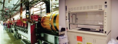
Because of the different areas of science, there are many different types of science labs that
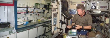
each include different scientific equipment. For example, a physics lab might contain a particle accelerator, in which the particles that make up atoms are studied. A chemistry or biology lab most likely contains a fume hood where substances with poisonous fumes can be worked. A particle accelerator and a fume hood are both shown in Figure 1.28. Despite the great differences among labs, some features are common in them.
Most labs have workbenches or countertops at which the scientist may sit or stand to do work comfortably. This is important because scientists can spend all day working in the lab. A scientist usually records an experiment’s progress in a lab notebook, but modern labs almost always contain a computer for data collection and analysis. In many labs computers are also used for lab simulations (modeling or imitating an experiment or a natural process), and for presenting results in the form of graphs or tables.
Lab Equipment
Lab techniques include the procedures used in science to carry out an experiment. Lab tech- niques follow scientific methods, and while some of them involve the use of simple laboratory equipment such as glassware (shown on the shelves in Figure 1.27), others use more complex
and expensive equipment such as electrical and computerized machines such as the particle accelerator shown in Figure 1.28, or use expensive supplies.
Equipment commonly found in a biology labs include microscopes, weighing scales or bal- ances, water baths, glassware (such as test tubes, flasks, and beakers), Bunsen burners, tongs, pipettes shown in Figure 1.29, chemical reagents, lab coats, goggles, and biohazard waste containers.

µL, which is smaller than a drop of water. (16)
Light Microscopes
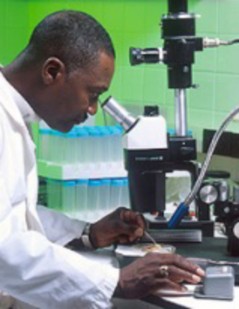
Microscopes are instruments used to view objects that are too small to be seen by the naked eye. Optical microscopes, such as the one shown in Figure 1.30, use visible light and lenses to magnify objects. They are the simplest and most widely used type of microscopes. Compound microscopes are optical microscopes which have a series of lenses, and have uses in many fields of science, particularly biology and geology. The scientist in Figure 1.31 is looking through a compound light microscope that is fitted with a digital camera.
Resolution is a measure of the clarity of an image; it is the minimum distance two points can be separated and still be distinguished as two separate points. Because light beams have a physical size, which is described in wavelengths, it is difficult to see an object that is about the same size or smaller than the wavelength of light. Objects smaller than about 0.2 micrometers appear fuzzy, and objects below that size cannot be seen.
Magnification involves enlarging the image of an object so that it appears much bigger than its actual size. Magnification also refers to the number of times an object is magnified. For example, a lens that magnifies 100X, magnifies an object 100 times larger than its actual size. Light microscopes have three objective lenses that have different magnifications, as
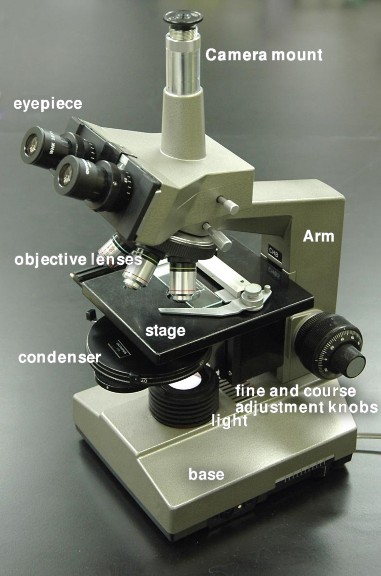
shown in Figure 1.32. The ocular lens has a magnification of 10X, so a 100X objective lens and the ocular lens together will magnify an object by 1000X.

Figure 1.32: Objective lenses of a light microscope. (48)
Visible light has wavelengths of 400 to 700 nanometers, which is larger than many objects of interest such as the insides of cells. Scientists use different types of microscopes in order to get better resolution and magnification of objects that are smaller than the wavelength of visible light. Objects that are to be viewed under an electron microscope may need to be specially prepared to make them suitable for magnification.
Electron Microscopes
Electron microscopes use electrons instead of photons (light), because electrons have a much shorter wavelength than photons and thus allow a researcher to see things at very high magnification, far higher than an optical microscope can possibly magnify.
There are two general types of electron microscopes: the Transmission Electron Microscope that shoots electrons through the sample and measures how the electron beam changes because it is scattered in the sample, and the Scanning Electron Microscope that scans an electron beam over the surface of an object and measures how many electrons are scattered back.
Transmission electron microscopy (TEM) is an imaging method in which a beam of electrons is passed through a specimen. An image is formed on photographic film or a fluorescent screen by the electrons that scatter when passing through the object. TEM images show the inside of the object.
The scanning electron microscope (SEM) is a type of electron microscope capable of producing high-resolution images of a sample surface. Due to the manner in which the image is created, SEM images have a characteristic three-dimensional appearance and are useful for judging the surface structure of the sample. Sometimes objects need to be specially prepared to make them better suited for imaging under the scanning electron microscope, as shown with the insect in Figure 1.33.
Electron microscopes work under low pressures and usually in a vacuum chamber to avoid scattering the electrons in the gas. This makes the microscopes considerably larger and more expensive than optical microscopes. The different types of images from the two electron microscopes are shown in Figure 1.34.
Aseptic Technique
In the microbiology lab, aseptic technique refers to the procedures that are carried out under sterile conditions. Scientists who study microbes are called microbiologists. Micro- biologists must carry out their lab work using the aseptic technique to prevent microbial contamination of themselves, contamination of the environment they are working in, in- cluding work surfaces or equipment, and contamination of the sample they are working on. Bacteria live on just about every surface on Earth, so if a scientist wants to grow a particular type of bacterium in the lab, he or she needs to be able to sterilize their equipment to prevent contamination by other bacteria or microorganisms. The aseptic technique is also used in medicine, where it is important to keep the human body free of contamination.
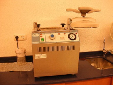
Aseptic technique is used whenever bacteria or other microbes are transferred between nu- trient media or in the preparation of the nutrient media. Some equipment that is used in the aseptic technique include a Bunsen burner, an autoclave (Figure 1.35), hand and surface sanitizers, neoprene gloves, and a fume hood.
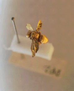
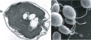
Figure 1.34: SEM and TEM images of the algae Chlamydomonas. The SEM image, shown at the right, is a three-dimensional image of the surface of the organism, whereas the TEM image is a two-dimensional image of the interior of the organism. (32)
Students of microbiology are taught the principles of aseptic technique by hands-on labora- tory practice. Practice is essential in learning how to handle the lab tools without contami- nating them.
Scientific Models
Scientific models are representations of reality. To describe particular parts of a phenomenon, or the interactions among a set of phenomena, it is sometimes helpful to develop a model of the phenomenon. For instance, a scale model of a house or of a solar system is clearly not an actual house or an actual solar system; the parts of an actual house or an actual solar system represented by a scale model are, only in limited ways, representative of the actual objects.
Scientific modeling is the process of making abstract models of natural phenomena. An abstract model is a theoretical construct that represents something. Models are developed to allow reasoning within a simplified framework that is similar to the phenomena being investigated. The simplified model may assume certain things that are known to be incom- plete in some details. Such assumptions can be useful in that they simplify the model, while at the same time, allowing the development of acceptably accurate solutions. These models play an important role in developing scientific theories.
A simulation is a model that runs over time. A simulation brings a model to life and
shows how a particular object or phenomenon will behave. It is useful for testing, analysis or training where real-world systems or concepts can be represented by a model. For the scientist, a model also provides a way for calculations to be expanded to explore what might happen in different situations. This method often takes the form of models that can be programmed into computers. The scientist controls the basic assumptions about the variables in the model, and the computer runs the simulation, eventually coming to a complicated answer.
Examples of models include:
Computer models
Weather forecast models
Molecular models
Climate models
Ecosystem models
Geologic models
One of the main aims of scientific modeling is to allow researchers to quantify their obser- vations about the world. In this way, researchers hope to see new things that may have escaped the notice of other researchers. There are many techniques that model builders use which allow us to discover things about a phenomenon that may not be obvious to everyone.
The National Weather Service Enhanced Radar Images web site (http://radar. weather.gov/) is an excellent example of a simulation. The site exhibits current weather forecasts across the United States.
Evaluating Models
A person who builds a model must be able to recognize whether a model reflects reality. They must also be able to identify and work with differences between actual data and theory.
A model is evaluated mostly by how it reflects past observations of the phenomenon. Any model that is not consistent with reproducible observations must be modified or rejected. However, a fit to observed data alone is not enough for a model to be accepted as valid. Other factors important in evaluating a model include:
Its ability to explain past observations
Its ability to predict future observations
Its ability to control events
The cost of its use, especially when used with other models
Ease of use and how it looks
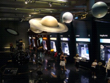
Some examples of the different types of models that are used by science are shown in Figures
1.37 and 1.38.
Theories as ”Models”
Theories are constructed in order to explain, predict and understand phenomena. This could include the movement of planets, weather patterns, or the behavior of animals, for example. In many instances we are constructing models of reality. A theory makes generalizations about observations and is made up of a related set of ideas and models. The important difference between theories and models is that the first is explanatory as well as descriptive, while the second is only descriptive and predictive in a much more limited sense.
Lab Safety
In some laboratories, conditions are no more dangerous than in any other room. In many labs, though, additional hazards are present. Laboratory hazards are as varied as the subjects of study in laboratories, and might include poisons, infectious agents, flammable, explosive, or radioactive materials, moving machinery, extreme temperatures, or high voltage. The hazard symbols for corrosive, explosive, and flammable substances are shown in Figure 1.39. In laboratories where conditions might be dangerous, safety precautions are important. La

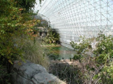
Figure 1.38: Biosphere 2 is an example of a very large three-dimensional model which biol- ogists built to attempt to recreate a self-sustaining biome. To learn more about biomes and ecosystems, go to the Biomes, Ecosystems and Communities chapter. (5)

Figure 1.39: The hazard symbols for corrosive, explosive, and flammable substances. (40)

safety rules minimize a person’s risk of getting hurt, and safety equipment is used to protect the lab user from injury or to help in responding to an emergency.
Some safety equipment that you might find in a biology lab includes:
Sharps Container A container that is filled with used medical needles and other sharp instruments such as blades, shown in Figure 1.40. Needles or other sharp items that have been used are dropped into the container without touching the outside of the container. Objects should never be pushed or forced into the container, as damage to the container or injuries may result.
Laminar Flow Cabinet A carefully enclosed bench designed to prevent contamination of biological samples. Air is drawn through a fine filter and blown in a very smooth, laminar (streamlined) flow towards the user. The cabinet is usually made of stainless steel with no gaps or joints where microorganisms might collect.
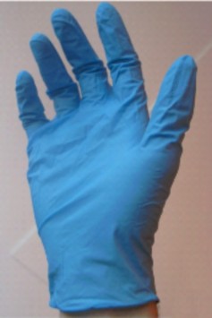
Gloves Due to possible allergic reactions to latex, latex gloves are not recommended for lab use. Instead, vinyl or nitrile gloves, shown in Figure 1.41, are often used. Gloves protect the wearers hands and skin from getting contaminated by microorganisms or stained or irritated by chemicals.
Lab Coat A knee-length overcoat that is usually worn while working in the lab. The coat
helps to protect the researcher’s clothes from splashes or contamination. The garment is made from white cotton or linen to allow it to be washed at high temperature and make it easy to see if it is clean.
Safe Laboratory Practice
Safety precautions are in place to help prevent accidents. Always wear personal protective equipment such as goggles and gloves when recommended to do so by your teacher.
Tell your teacher immediately if an accident happens.
The production of aerosols due to poor technique such as squirting the last drop out of pipettes, and the spread of contamination due to spills is completely avoidable and especially important if you are handling infectious material or chemicals.
Wear enclosed toe shoes, instead of sandals or flip flops, or thongs. Your feet and toes could easily get hurt or broken or if you dropped something. (Figure 1.42)
Do not wear loose, floppy clothes in the lab; they can get caught in or knock over equipment, causing an accident.
If you have long hair, tie it up for the same reasons listed above.
Do not eat or drink in the lab.
Do not use cell phones in the lab, even if you are only sending a text message. You can easily contaminate your phone with whatever you have been working with. Consider where your hands have been, and where your face will be the next time you talk on the phone.
Sweep up broken glass immediately and dispose in a designated area or container, or notify your teacher.
Always listen carefully to your teacher’s instructions.

Accidents
In the case of an accident, it is important to begin by telling your teacher and to know where to find safety equipment.
Some common safety equipment in a school lab:
Fire Extinguishers
Fire Blanket
Eye-Wash Fountain (Figure 1.43)
First-Aid Kit
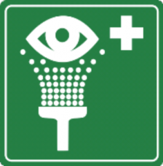
Figure 1.43: Symbol for the eyewash fountain. (51)
Through the first three lessons, we have discussed what science is and how science is done. Now we need to turn our attention to Biology. Biology is the study of life. As the ‘study of life,’ a knowledge of biology is an extremely important aspect of your education. Biology includes the identification and analysis of characteristics common to all living organisms. What is known about biology is discovered or identified through the same processes as all other sciences, including the scientific method and peer review process.
Lesson Summary
The measurements that scientists use are based on the International System of Units (SI), which is form of the metric system. Based on multiples of ten, It is the world’s most widely used system of units, both in science and business.
One important use for mathematics in science is the role it plays in expressing scientific models. Statistics allow scientists to assess the reliability and range of differences in experimental results.
Light microscopes use visible light and lenses to magnify objects. They are the simplest and most widely used type of microscopes. Electron microscopes use electrons instead of photons (light), because electrons have a much shorter wavelength than photons and therefore allow a researcher to see things at very high magnification, that greatly exceeds what an optical microscope can possibly magnify. Electron microscopes are larger and more expensive than light microscopes.
Equipment commonly found in a biology labs include microscopes, weighing scales or balances, water baths, glassware (such as test tubes, flasks, and beakers), Bunsen burners, tongs, pipettes, chemical reagents, lab coats, goggles, and biohazard waste containers.
Always wear personal protective equipment such as goggles and gloves, wear enclosed shoes, and do not eat or drink in the lab.
Review Questions
Further Reading / Supplemental Links
Which one of the following units of measurement would be the most appropriate in determining the mass of a banana? Kilograms, micrograms, or grams.
Identify the type of microscope that is most common in laboratories.
Contrast microscope magnification and resolution.
If an objective lens magnifies an object by 45×, and the optical lens magnifies by 10×. By how much will the object be magnified to the viewer?
Which object is larger? An object with a diameter of 1500 micrometers (µm) or an object with a diameter of 15 millimeters (mm)?
Why is it important that scientists use common units of measurement?
Name three pieces of safety equipment that you should wear while carrying out an investigation in the lab.
What should you first do if an accident happens in the lab?
If you saw this hazard sign on a chemical container, what do you think it might mean?

How are computer models similar to the real world, and how do they differ?
http://www.chem.unl.edu/safety/hslabcon.html
http://en.wikibooks.org/wiki/Nanotechnology/Electron_microscopy
Vocabulary
aseptic technique Laboratory procedures that are carried out under sterile conditions.
compound microscope An optical microscopes that has a series of lenses, and have uses in many fields of science, particularly biology and geology.
electron microscope A microscope that uses electrons instead of light; allow a researcher to see things at very high magnification, far higher than an optical microscope can possibly magnify.
International System of Units (SI) The measurements that scientists use; a form of the metric system.
lab coat A knee-length overcoat that is usually worn while working in the lab; helps to protect the researcher’s clothes from splashes or contamination.
laboratory A place that has controlled conditions in which scientific research, experiments, and measurement may be carried out.
lab techniques The procedures used in science to carry out an experiment.
magnification Enlarging an image of an object so that it appears much bigger than its actual size; also refers to the number of times an object is magnified.
microscopes Instruments used to view objects that are too small to be seen by the naked eye.
model A physical, mathematical, or logical representation of a system, phenomenon, or process; allow scientists to investigate a phenomenon in a controlled way.
optical microscope A microscope that uses visible light and lenses to magnify objects.
resolution A measure of the clarity of an image; it is the minimum distance two points can be separated and still be distinguished as two separate points.
scanning electron microscope (SEM) Electron microscope that scans an electron beam over the surface of an object and measures how many electrons are scattered back.
scientific modeling The process of making abstract models of natural phenomena.
simulation A model that runs over time; brings a model to life and shows how a particular object or phenomenon will behave.
stereo microscope A light microscope with two ocular lenses.
transmission electron microscope (TEM) Electron microscope that shoots electrons through the sample and measures how the electron beam changes because it is scattered in the sample.
Points to Consider
Consider how much more difficult it would be to carry out investigations without the use of computers, and the types of models that have developed due to the development of computers.
Consider reasons why eating and drinking are not allowed in the lab.
What additional ethical considerations would there be if you were working with living organisms in the lab, such as mice, rats, or other mammals?
- Log in or register to post comments
- Email this page