Lesson 22.3: Respiratory System
Lesson 22.3: Respiratory System
Lesson Objectives
Distinguish between external and internal respiration.
Identify the structures of the respiratory system.
Outline the process of inhalation.
Describe how carbon dioxide is carried in the blood.
Compare the causes of emphysema and asthma.
Introduction
Have you ever wondered what it would be like to have gills? You would breathe and look very different from the rest of us, but they would be great for swimming and diving! Despite such differences, the main functions of lungs and gills are the same: to obtain oxygen, and to release carbon dioxide.
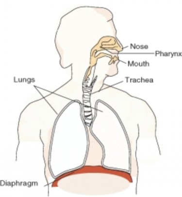
The human respiratory system brings oxygen, O2, into the body and releases carbon dioxide, CO2, into the atmosphere. Oxygen is drawn in through the respiratory tract, which is shown in Figure 22.34, and is then delivered to the blood. This process is called external respiration. The exchange of gases between the blood and the cells of the body is called internal respiration.
Comparing ”Cellular Respiration” and ”Respiration”
Respiration is the transport of oxygen from the outside air to the cells of the body, and the transport of carbon dioxide in the opposite direction. This is in contrast to the biochemical definition of respiration, which refers to cellular respiration. Cellular respiration is the metabolic process by which an organism obtains energy by reacting oxygen with glucose to give water, carbon dioxide and ATP (energy). Although respiration is necessary to sustain cellular respiration and thus life in animals, the processes are very different. Cellular respiration takes place in individual cells of the animal, while respiration involves the transport of metabolites between the organism and external environment.
Structures of the Respiratory System
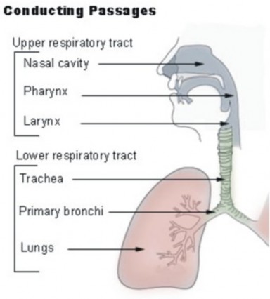
The nose and nasal cavity filter, warm, and moisten the inhaled air. The nose hairs and mucus produced by the epithelial cells in the nose catch airborne particles and prevent them from reaching the lungs.
Behind the nasal cavity, air next passes through the pharynx, a long tube that is shared with the digestive system. Both food and air pass through the pharynx. A flap of connective tissue called the epiglottis closes over the trachea when food is swallowed to prevent choking or inhaling food. In humans the pharynx is important in vocalization
The larynx, also called the voicebox, is found just below the point at which the pharynx splits into the trachea and the esophagus, shown in Figure 22.35. The voice is generated in the larynx. Air from the lungs is needed for the vocal folds to produce speech.
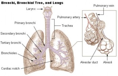
The trachea, or wind pipe, is a long tube that leads down to the chest where it divides into the right and left bronchi in the lungs. The bronchi branch out into smaller bronchioles, which are the first airway passages that do not contain cartilage. The bronchioles lead into the alveoli, which are the multi-lobed sacs in which most of the gas exchange occurs.
The Journey of a Breath of Air
In air-breathing vertebrates such as humans, respiration of oxygen includes four stages:
Ventilation: From the Air to the Alveoli
Air enters the body through the nose, is warmed, filtered, and passed through the nasal cavity. Air passes the pharynx (which has the epiglottis that prevents food from entering the trachea). The upper part of the trachea contains the larynx. The vocal cords are two bands of tissue that extend across the opening of the larynx. After passing the larynx, the air moves into the trachea. The trachea is a long tube that divides into two smaller tubes called bronchi which lead into each lung, shown in Figure 22.35. Bronchi are reinforced to prevent their collapse and are lined with ciliated epithelium and mucus-producing cells.
Bronchi branch into smaller and smaller tubes called bronchioles. Bronchioles end in grape- like clusters called alveoli. Alveoli are surrounded by a network of thin-walled capillaries, shown in Figure 22.36.
Breathing in, or inhaling, is usually an active movement, contraction of the diaphragm muscles uses ATP. The diaphragm is a muscle that is found below the lungs (shown in Figure 22.34). Contraction of the diaphragm causes the volume of the chest cavity to increase, and the air pressure within the lungs to decrease. The pressure difference causes air to rush into the lungs. Relaxation of the diaphragm causes the lungs to recoil and air is pushed out of the lungs. Breathing out, or exhaling, is normally a passive process powered by the elastic recoil of the chest, similar to letting the air out of a balloon.
Pulmonary Gas Exchange: From the Alveoli into the Pulmonary Capillaries
Breathing is only part of the process of delivering oxygen to where it is needed in the body. The process of gas exchange occurs in the alveoli by diffusion of gases between the alveoli and the blood passing in the lung capillaries, as shown in Figure 22.37. Recall that diffusion is the movement of substances from an area of higher concentration to an area of lower concentration. The difference between the high concentration of O2 in the alveoli and the low O2 concentration of the blood in the capillaries is enough to cause O2 molecules to diffuse across the thin walls of the alveoli and capillaries and into the blood. CO2 moves out of the blood and into the alveoli in a similar way. The greater the concentration difference, the greater the rate of diffusion.
Breathing also results in loss of water from the body. Exhaled air has a relative humidity
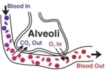
Figure 22.37: Gas exchange happens in the lungs through diffusion. Deoxygenated blood has a high concentration of CO2 and a low O2 concentration. CO2 moves out of the blood and into the alveoli, where the concentration of CO2 is lower. Likewise, O2 moves from an area of higher concentration (the alveoli), to an area of lower concentration (the blood). (29)
of 100 percent because of the diffusion of water that from the moist surface of the breathing passages and the alveoli into the warm exhaled air.
In the lungs, oxygen is transported across the thin membranes of the alveoli and the border of the capillary and attracted to the hemoglobin molecule within the red blood cell.
After leaving the lungs, the oxygenated blood returns to the heart to be pumped through the aorta and around the body. The oxygenated blood travels through the aorta, to the smaller arteries, arterioles, and finally to the peripheral capillaries where gas exchange occurs.
Peripheral Gas Exchange: From Capillaries into Cells, and from Cells into Capillaries
The oxygen concentration in body cells is low, while the blood that leaves the lungs is 97 percent saturated with oxygen. So, oxygen diffuses from the blood into the body cells when it reaches the peripheral capillaries (the capillaries in the systemic circulation).
Carbon dioxide concentration in metabolically active cells is much greater than in capillaries, so carbon dioxide diffuses from the cells into the capillaries. Most of the carbon dioxide (about 70 percent) in the blood is in the form of bicarbonate (HCO3-). A small amount of carbon dioxide dissolves in the water in the plasma to form carbonic acid (H2CO3). Carbonic acid and bicarbonate play an important role in regulating the pH of the body.
In order to remove CO2 from the body, the bicarbonate is picked up by a red blood cell, and is again turned in to carbonic acid. A water molecule (H2O) is then taken away from the carbonic acid, and the remaining CO2 molecule is expelled from the red blood cells and into the alveoli where it is exhaled. The following equation shows this process:
HCO3- + H+ H2CO3 CO2 + H2O
Gas exchange between your body and the environment occurs in the alveoli. The alveoli are lined with pulmonary capillaries, the walls of which are thin enough to permit the diffusion of gases. Inhaled oxygen diffuses into the pulmonary capillaries, where it binds to hemoglobin in the blood. Carbon dioxide diffuses in the opposite direction, from capillary blood to alveolar air. At this point, the pulmonary blood is oxygen-rich, and the lungs are primarily holding carbon dioxide. Exhalation follows, thereby ridding the body of the carbon dioxide and completing the cycle of respiration.
Gas Exchange and Homeostasis
The equilibrium between carbon dioxide and carbonic acid is very important for controlling the acidity of body fluids. As gas exchange occurs, the pH balance of the body is maintained as part of homeostasis. If proper respiration is interrupted two things can occur:
Control of Breathing by the Nervous System
Breathing is one of the few bodily functions which, within limits, can be controlled both consciously and unconsciously. Conscious attention to breathing is common in activities such as yoga, swimming, and karate. In speech or vocal training, a person learns to discipline his or her breathing for purposes other than life support.
Muscular contraction and relaxation controls the rate of expansion and constriction of the lungs. These muscles are controlled by the autonomic nervous system from the parts of the brainstem that control breathing: the medulla and the pons. This area of the brainstem forms the respiration regulatory center. When carbon dioxide levels increase in the blood (in the form of carbonic acid), such as during exercise, the pH level of the blood drops. This causes the medulla to send nerve impulses to the diaphragm and the muscles between the ribs, causing them to contract and increase the rate of breathing. This automatic control of respiration can be impaired in premature babies, or by drugs or disease.
Without breathing, the body’s oxygen levels drop dangerously low within minutes, leading to permanent brain damage followed by death. It is not possible for a healthy person to voluntarily stop breathing indefinitely. If we do not inhale, the level of carbon dioxide builds up in our blood and we experience great air hunger. Eventually, not breathing leads to a loss of consciousness at which time the autonomic nervous system takes control and initiates breathing.
Inhalation
Inhalation is started by the diaphragm and supported by the external intercostal muscles (the muscles that are between the ribs). It is an active process that needs ATP. When the diaphragm contracts, the ribcage expands and the contents of the abdomen are moved downward. This results in a larger thoracic (chest) volume, which in turn causes a decrease in air pressure inside the lungs. As the pressure in the chest falls, air from outside the body moves into the respiratory system. Normal resting respirations are 10 to 18 breaths per minute. During an average breath, an adult will exchange from 500 ml to 700 ml of air. The average breath capacity of a person is called lung volume, or tidal volume.
Exhalation
Exhalation is generally a passive process, however active, or forced, exhalation is carried out by the abdominal and the internal intercostal muscles. The lungs have a natural elasticity and as they recoil from the stretch of inhalation, air flows out of the lungs until the pressures in the chest and the atmosphere reach equilibrium. During forced exhalation, as when blowing out a candle, expiratory muscles including the abdominal muscles and internal intercostal muscles generate pressure in the chest and abdomen, which forces air out of the lungs.
Homeostatic Imbalances of the Respiratory System: Dis- eases and Disorders
Respiratory disease is the term for diseases of the lung, bronchial tubes, trachea and throat. These diseases range from mild, such as a cold, to being possibly life-threatening, such as bacterial pneumonia.
Respiratory diseases can be grouped as either obstructive (conditions which lower the rate of the airflow into and out of the lungs, such as in asthma) or restrictive (conditions that cause a reduction in the functional volume of the lungs, such as emphysema.)
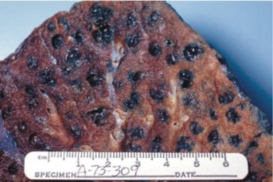
Emphysema is a chronic lung disease caused by loss of elasticity of the lung tissue. The destruction of elastic structures that support the alveoli and the capillaries that feed the alveoli cause them to become hard and stiff. Eventually the walls of the alveoli break down and the alveoli become larger. The amount of oxygen that can enter the blood with each breath is reduced because the large alveoli cannot function efficiently; much of the oxygen that gets into the large alveoli cannot be absorbed into the blood so the oxygen is unused.
Symptoms include shortness of breath on exertion (usually when climbing stairs or a hill, and later at rest), and an expanded chest. Damage to the alveoli, which can be seen in Figure 22.38, is irreversible. Smoking is a leading cause of emphysema.
Bronchitis is an inflammation of the bronchi. Acute bronchitis is usually caused by viruses or bacteria and may last several days or weeks. Acute bronchitis is characterized by cough and phlegm (mucus) production. Symptoms are related to the inflammation of the airways and phlegm production, and include shortness of breath and wheezing. Chronic bronchitis is not necessarily caused by infection and is generally part of a syndrome called chronic ob- structive pulmonary disease (COPD). Chronic bronchitis is defined clinically as a persistent cough that produces phlegm and mucus, for at least three months in two consecutive years.
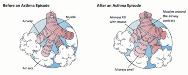
Asthma is a chronic illness in which the airways narrow and becomes inflamed, as shown in Figure 22.39. Excessive amounts of mucus are also made by the lungs. Asthma often happens in response to one or more triggers. It may be triggered by exposure to an allergen such as mold, dust, or pet hair. It can also be caused by cold air, warm air, moist air, exercise, or emotional stress. In children, the most common triggers are viral illnesses such as those that cause the common cold. This airway narrowing causes symptoms such as wheezing, shortness of breath, chest tightness, and coughing. Some people with asthma, especially children, can become very frightened by the symptoms, which may cause even more breathing distress. Between asthma attacks, most patients feel well but can have mild symptoms and may remain short of breath after exercise for longer periods of time than a person who does not have asthma. The symptoms of asthma, which can range from mild to life threatening, can usually be controlled with a combination of medicines and environmental changes.
Public attention in the developed world has recently focused on asthma because of the increasing numbers of cases, affecting up to one in four children who live in cities.
Pneumonia is an illness in which the alveoli become inflamed and flooded with fluid. Ef- fective gas exchange cannot happen across the alveoli membranes. Pneumonia can result from a variety of causes, including infection with bacteria, viruses, fungi, or parasites, and chemical or physical injury to the lungs. Symptoms of pneumonia include cough, chest pain, fever, and difficulty in breathing. Treatment depends on the cause of pneumonia; bacterial pneumonia is treated with antibiotics.
Pneumonia is a common illness which occurs in all age groups, and is a leading cause of death among the elderly and people who are chronically and terminally ill. Vaccines to prevent certain types of pneumonia are available.
Tuberculosis (TB) is a common and deadly infectious disease caused by a type of bacteria called Mycobacterium tuberculosis. TB most commonly attacks the lungs (as pulmonary TB) but can also affect the central nervous system, the lymphatic system, the circulatory system, the genitourinary system, bones, joints and even the skin.
Over one-third of the world’s population has been exposed to the TB bacterium. Not ev- eryone infected develops the disease, so TB infection without symptoms (called a latent infection) is most common. However, one in ten latent infections will progress to active TB disease, which, if left untreated, kills more than half of its victims.
The rise in HIV infections and the neglect of TB control programs have led to an increase in cases of tuberculosis. The development of drug-resistant strains has also contributed to this new epidemic. For example, between 2000 and 2004, about 20 percent of TB cases were resistant to standard antibiotic treatments. TB incidence varies widely, even in neighboring countries, apparently because of differences in health care system standards. A TB vaccine, called Bacille Calmette-Guérin (BCG), is available to people in some countries. The BCG is prepared from a strain of weakened live mycobacterium, which has lost its virulence in humans. The effectiveness of the BCG is a matter of debate among researchers, and the governments in some countries, including the United States, do not require people to get the BCG vaccination.
Lung cancer is a disease where epithelial (internal lining) tissue in the lung grows out of control. This leads to invasion of nearby tissue and growth of the tumor beyond the lungs. Lung cancer, which is the most common cause of cancer-related death in men and the second most common in women, is responsible for 1.3 million deaths worldwide every year The most common symptoms are shortness of breath, coughing (including coughing up blood), and weight loss.
The most common cause of lung cancer is exposure to tobacco smoke. The occurrence of lung cancer in non-smokers, who account for less than 10 percent of cases, appears to be due to a combination of genetic factors. Radon gas, asbestos, and air pollution may also contribute to lung cancer.
Asbestos is a mineral that was once used as a fire retardant in buildings and electrical wiring. The inhalation of asbestos fibers can cause a variety of lung diseases, including lung cancer. Tobacco smoking and exposure to asbestos greatly increase a person’s chance of developing lung cancer.
Lesson Summary
Ventilation from the atmosphere into the alveoli of the lungs.
Pulmonary gas exchange from the alveoli into the pulmonary capillaries.
Gas transport from the pulmonary capillaries through the circulation to the peripheral capillaries in the organs.
Peripheral gas exchange from the tissue capillaries into the cells and mitochondria.
Respiratory acidosis, in which arterial blood contains too much carbon dioxide, causing a drop in blood pH.
Respiratory alkalosis results from increased respiration (or hyperventilation) which causes a drop in the amount of carbon dioxide in the blood plasma. The drop in carbon dioxide concentration causes the blood pH to rise.
The main functions of lungs are to obtain oxygen, and to release carbon dioxide. Oxygen is drawn in through the respiratory tract and is then delivered to the blood in a process called external respiration. The exchange of gases between the blood and the cells of the body is celled internal respiration.
The structures of the respiratory systems include the nose and nasal cavity, the phar- ynx, the larynx, ( also called the voicebox), the trachea (also called the wind pipe), the right and left bronchi in the lungs, and the bronchioles that end in the alveoli.
During inhalation, the diaphragm contracts, causing the volume of the chest cavity to increase. As a result, the air pressure within the lungs decreases. The pressure difference causes air to rush into the lungs. Relaxation of the diaphragm causes the lungs to recoil and air is pushed out of the lungs, which causes exhalation.
Most of the carbon dioxide (about 70 percent) in the blood is in the form of bicarbonate (HCO3-). A small amount of carbon dioxide dissolves in the water in the plasma to form carbonic acid (H2CO3). When CO2 enters the blood from body cells, it combines with water in the plasma to produce carbonic acid (H2CO3), which is then turned into bicarbonate (HCO3-). The bicarbonate is then picked up by a red blood cell and turned back in to carbonic acid. A water molecule (H2O) is then taken away from the carbonic acid, and the remaining CO2 molecule is expelled from the red blood cells and into the lungs.
Emphysema is a chronic lung disease caused by loss of elasticity of the lung tissue. The destruction of elastic structures that support the alveoli and the capillaries that feed the alveoli cause them to become hard and stiff. It is often caused by smoking. Asthma is also a chronic condition, which is often triggered by such things as exposure to an allergen, cold air, warm air, moist air, exercise, or emotional stress. The airways can constrict and become inflamed, and an excessive amount of mucus is produced. Airway narrowing causes symptoms such as wheezing, shortness of breath, chest tightness, and coughing.
Review Questions
Further Reading / Supplemental Links
Identify the respiratory structures through which air flows.
How is the diaphragm involved in breathing?
Compare respiration and cellular respiration.
Outline how most carbon dioxide is carried in the blood.
Why is it important for a pregnant woman to know her Rhesus blood type, and the Rh blood type of the father of her baby?
What is the difference between internal and external respiration?
What happens during an asthma attack?
Outline how emphysema affects the absorption of oxygen.
Where does the exchange of oxygen occur in the lungs?
What factors regulate breathing rate?
http://www.estrellamountain.edu/faculty/farabee/biobk/BioBookRESPSYS.html
Vocabulary
alveoli Multi-lobed sacs in which most of the gas exchange occurs.
asthma A chronic illness in which the airways narrow and becomes inflamed.
bronchitis An inflammation of the bronchi.
diaphragm A muscle that is found below the lungs; contraction of the diaphragm causes the volume of the chest cavity to increase, and the air pressure within the lungs to decrease.
emphysema A chronic lung disease caused by loss of elasticity of the lung tissue.
external respiration Process in which oxygen is drawn in through the respiratory tract and is then delivered to the blood.
gas exchange The diffusion of gases between the alveoli and the blood passing in the lung capillaries; also the diffusion of gases from capillaries into cells, and from cells into capillaries throughout the body (peripheral gas exchange).
internal respiration The exchange of gases between the blood and the cells of the body.
larynx Found just below the point at which the pharynx splits into the trachea and the esophagus; also called the voice box.
lung cancer A disease where epithelial (internal lining) tissue in the lung grows out of control; leads to invasion of nearby tissue and growth of the tumor beyond the lungs.
lung volume (tidal volume) The average breath capacity of a person.
obstructive Conditions which lower the rate of the airflow into and out of the lungs, such as in asthma.
pharynx A long tube that is shared with the digestive system; both food and air pass through the pharynx.
pneumonia An illness in which the alveoli become inflamed and flooded with fluid.
respiration The transport of oxygen from the outside air to the cells of the body, and the transport of carbon dioxide in the opposite direction.
respiratory acidosis Condition in which arterial blood contains too much carbon dioxide, causing a drop in blood pH.
respiratory alkalosis Condition which results from increased respiration (or hyperventi- lation) which causes a drop in the amount of carbon dioxide in the blood plasma; the drop in carbon dioxide concentration causes the blood pH to rise.
respiratory disease The term for diseases of the lung, bronchial tubes, trachea and throat.
restrictive Conditions that cause a reduction in the functional volume of the lungs, such as emphysema.
trachea A long tube that leads down to the chest where it divides into the right and left bronchi in the lungs; also called the windpipe.
tuberculosis (TB) A common and deadly infectious disease caused by a type of bacteria called Mycobacterium tuberculosis; most commonly attacks the lungs, but can also affect the central nervous system, the lymphatic system, the circulatory system, the genitourinary system, bones, joints and even the skin.
Points to Consider
How might the amount of oxygen in the air affect your respiratory and circulatory systems?
Can you identify any structures that are part of both the respiratory and digestive systems?
Image Sources
Drs. Noguchi, Rodgers, and Schechter of NIDDK.
http://en.wikipedia.org/wiki/Image:Sicklecells.jpg. Public Domain.
USFG. http://commons.wikimedia.org/wiki/Image:Asthma_before-after.png. Public Domain.
http://visualsonline.cancer.gov/details.cfm?imageid=2129. Public Domain.
A. Rad. http://commons.wikimedia.org/wiki/File:Coombs_test_schematic.png. CC-BY-SA.
http://commons.wikimedia.org/wiki/Image: Heart_left_lateral_coronaries_diagram.svg. CC-BY-2.5.
USFG. http://commons.wikimedia.org/wiki/Image:Illu_lymph_capillary.png. Public Domain.
http://commons.wikimedia.org/wiki/Image:Macrophage.jpg. CC-BY-SA-2.0.
USFG. Internal structure of a vein.. Public Domain.
http://en.wikipedia.org/wiki/Image:Bundleofhis.png. Public Domain.
Data from Braunwald’s Heart Disease: A Textbook of Cardiovascular Medicine. 7th ed.. W.B. Saunders Company; Philadelphia 2004..http://commons.wikimedia.org/wiki/Image:CVD_Praevalenz_US.png.
CC-BY-SA.
http://commons.wikimedia.org/wiki/Image:Sphygmomanometer.jpg. GNU-FDL, Public Domain.
http://www.flickr.com/photos/johnjoh/349825408/. CC-BY-SA, CC-BY-SA.
J. Heuser. http://en.wikipedia.org/wiki/Image:AMI_scheme.png.
http://commons.wikimedia.org/wiki/Image:Red_White_Blood_cells.jpg. Public Domain.
http://training.seer.cancer.gov/anatomy/cardiovascular/heart/ structure.html. Public Domain.
The structure of an artery wall.. Public Domain.
Apers0n. http://en.wikipedia.org/wiki/Image:Bedside_card.jpg. CC-BY.
//training.seer.cancer.gov/anatomy/cardiovascular/blood/pathways.html. Public Domain.
The direction of blood flow through the heart.. GNU-FDL.
http://commons.wikimedia.org/wiki/Image:Grafik_blutkreislauf.jpg. CC-BY-SA-2.5.
http://commons.wikimedia.org/wiki/Image: Heart_coronary_artery_lesion.jpg. CC-BY-2.5.
MesserWoland. http://commons.wikimedia.org/wiki/Image:Illu_blood_components.svg. CC-BY-SA.
USFG. Lymphatic system. Public Domain.
Center for Disease Control. Public Domain.
//training.seer.cancer.gov/anatomy/respiratory/passages/bronchi.html. Public Domain.
Theresa Knott. http://commons.wikimedia.org/wiki/Image:Respiratory_system.svg. CC-BY-SA-2.5.
http://en.wikipedia.org/wiki/Image:Veincrosssection.png. Public Domain, GNU-FDL.
Mr TS88 Duel. http://en.wikipedia.org/wiki/Image:Alveoli.jpg. GFDL/CC-BY-2.5.
USFG. http://en.wikipedia.org/wiki/Image:Illu_conducting_passages.jpg. Public Domain.
http://en.wikipedia.org/wiki/Image:Heart_diastole.png. GNU-FDL.
CDC/Dr. Edwin P. Ewing, Jr. http://commons.wikimedia.org/wiki/Image: Centrilobular_emphysema_865_lores.jpg. Public Domain.
http://en.wikipedia.org/wiki/Image:Double_circulatory_system.jpg. Public Domain.
http://en.wikipedia.org/wiki/Image:Purkinje_fibers.jpg. CC-BY-SA.
http://commons.wikimedia.org/wiki/Image:Elephantiasis.png. CC-BY-2.5.
http://en.wikipedia.org/wiki/Image:Illu_capillary.jpg. Public Domain.
http://en.wikipedia.org/wiki/Image:Illu_pulmonary_circuit.jpg. Public Domain.
http://commons.wikimedia.org/wiki/Image:Blood_smear.jpg. GNU-FDL.
http://commons.wikimedia.org/wiki/Image:Mediastinum_anatomy.jpg. CC-BY-2.5.
- Log in or register to post comments
- Email this page